PEROSSAL SOSITUTO OSSEO PER IL RIPRISTINO DEI DIFETTI OSSEI.
Dopo un accurato sbrigliamento chirurgico e in presenza di antibiotici sistemici e/o locali, può essere impiantato anche in aree infette o contaminate.
PerOssal® è un materiale sostitutivo osseo sintetico, biodegradabile e osteoconduttivo per la ricostruzione e il riempimento di difetti ossei. La sua esclusiva struttura microporosa assicura l’assorbimento uniforme di sostanze liquide (come gli antibiotici) e il loro rilascio controllato (eluizione) [1].
PerOssal® ha una struttura porosa che consente l’assorbimento sicuro di soluzioni acquose: 0,5 ml per 6 pellet e 4 ml per 50 pellet. Queste caratteristiche rendono PerOssal® uno scaffold ideale per il rilascio in loco di soluzione antibiotate.
COMPOSIZIONE
- 51.5% di idrossiapatite nano-cristallina
- 48,5% di solfato di calcio


PerOssal® è un pellet cilindrico di 6 mm x 6 mm, con una estremità sferica e una piatta. Sono disponibili confezioni da 1 × 6, 2 × 6 e 1 × 50 pellets . I pellets vengono confezionati in flaconcini, protetti da un doppio blister (confezione sterile interna ed esterna).
CARATTERISTICHE
- Nanocristallino/poroso: adatto come materiale di supporto per soluzioni acquose (come antibiotici).
- Personalizzabile: protezione antibiotica mirata e altamente efficace di Perossal, in base al singolo antibiogramma, con effetti collaterali sistemici minimi.
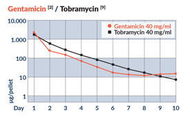
- Azione prolungata: dopo aver perfuso con antibiotici, Perossal rilascia a lungo termine (10/15 giorni) la protezione contro la colonizzazione di agenti patogeni batterici sensibili.
- Biodegradabile: completamente assorbibile entro 6 mesi [8, 10], in base alla dimensione del difetto, al sito di impianto e alla qualità dell’osso circostante. Nessuna seconda procedura richiesta per l’espianto.

RACCOMANDAZIONI DI DOSAGGIO ANTIBIOTICO*
* dosaggio raccomandato basato su risultati in vitro. Il medico curante è responsabile della decisione relativa al tipo e alla quantità dell’antibiotico utilizzato.
Devono essere considerate le controindicazioni dell’antibiotico applicato.
CONCENTRAZIONE
ANTIBIOTICO
Gentamicina
Tabromicina
Vancomicina
Rifampicina
40 mg/ml
40 mg/ml
50 mg/ml
60 mg/ml
RILASCIO IN VITRO DEGLI ANTIBIOTICI TESTATI CON PEROSSAL® PER UN PERIODO DI 10 GIORNI


INDICAZIONI
- PerOssal® è indicato per il riempimento o la ricostruzione di difetti ossei in caso di osso infetto o contaminato.
- PerOssal® è indicato, dopo un precedente intervento chirurgico, per la somministrazione sistemica e / o locale di antibiotici.
- PerOssal® può essere utilizzato come aumento dell’osso autologo [4].
AREE DI UTLIZZO


CHIRURGIA ORTOPEDICA
TRAUMATOLOGIA

CHIRURGIA MAXILLO-FACCIALE
CHIRURGIA SPINALE

CASO CLINICO
PAZIENTE DI 42 ANNI CON OSTEOMIELITE E FISTOLA DELLA TIBIA PROSSIMALE, 28 MESI DOPO L’OSTEOSINTESI CON PLACCA. [5]


Referenze
[1] Rauschmann et al. (2005), Nanocrystalline hydroxyapatite and calcium sulphate as biodegradable composite carrier material for local delivery of antibiotics in bone infections, Biomaterials. 26(15):2677-2684.
[2] Englert et al. (2007), Konduktives Knochenersatzmaterial mit variabler Antibiotikaversetzung
[Conductive bone substitute material with variable antibiotic delivery], Unfallchirurg. 110(5):408-413.
[3] Standardized bulk volume, data on file at OSARTIS GmbH.
[4] von Stechow and Rauschmann (2009), Effectiveness of combination use of antibiotic-loaded PerOssal® with spinal surgery in patients with spondylodiscitis, Eur Surg Res. 43(3):298-305.
[5] Kraus und Schnettler (2008), Gutachten bei Osteitis [Expert opinion in osteitis], in: Bericht über die Unfallmedizinische Tagung in Mainz am 8./9.11.2008, Deutsche Gesetzliche Unfallversicherung (Hrsg.), Heft 108, ISBN 3-88383-082-8.
[6] Fleege et al. (2012), Systemische und lokale Antibiotikatherapie bei konservativ und operativ behandelten Spondylodiszitiden [Systemic and local antibiotic therapy of conservative and operative treatment of spondylodiscitis], Orthopäde. 41(9):727-735.
[7] Fleege and Rauschmann (2013), Duration of antibiotic therapy after surgical treatment of nonspecific spondylodiscitis. Preliminary trends from a prospective randomized study short-term vs.
long-term antibiotic therapy, 32. Annual Meeting of the European Bone & Joint Infection Society 2013.
[8] Fleege et al. (2017), Antibiotikatherapie der pyogenen Spondylodiszitis bei Erwachsenen
[Antibiotic therapy of pyogenic spondylodiscitis in adults], Die Wirbelsäule. 01(4):265.
[9] Release kinetic data on file at OSARTIS GmbH.
[10] Visani et al. (2018), Treatment of chronic osteomyelitis with antibiotic-loaded bone void filler systems:
an experience with hydroxyapatites calcium-sulfate biomaterials, Acta Orthop Belg. 84(1):25-29.
